Assessing the immune state of the tumor and its microenvironment by imaging legumain and CD80
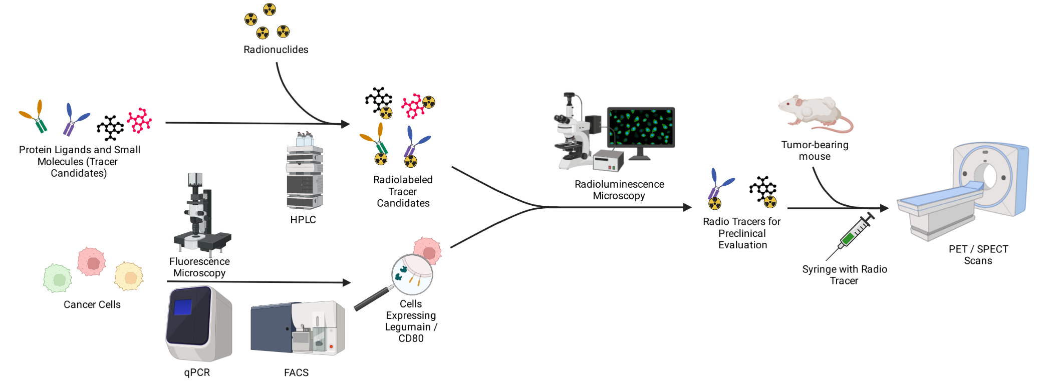
The majority of cancer therapies to date directly target malignant cells. However, the focus is increasingly being shifted to the tumor surroundings facilitating cancerous progression. Macrophages recruited to the tumor environment frequently express CD80 and legumain. Both CD80 and legumain represent promising targets for the diagnosis and monitoring of cancer immunotherapy. Our research efforts focus on the development of novel PET and SPECT tracers to image the presence of CD80 and legumain. We currently have multiple candidate small molecules and small protein ligands in the pipeline. To assess these candidates, cell based assays along mouse cancer models have been established by our group. In the course of model implementation, we employ a series of biological methods such as qPCR, FACS and fluorescence microscopy. To evaluate the radio tracer candidates, we apply radio synthesis and radio chelation followed by in vitro and in vivo techniques including radioluminescence microscopy and PET/SPECT scans.
Literature:
Taddio MF, Jaramillo CA, Runge P, Blanc A, Keller C, Talip Z, Béhé M, van der Meulen NP, Halin C, Schibli R, Krämer SD. In Vivo Imaging
of Local Inflammation: Monitoring LPS-Induced CD80/CD86 Upregulation by PET. Molecular imaging and biology. 2021 Apr;23(2):196-207.
