Monitoring progression and treatment efficiency of neurodegenerative and cardiovascular diseases with pet imaging
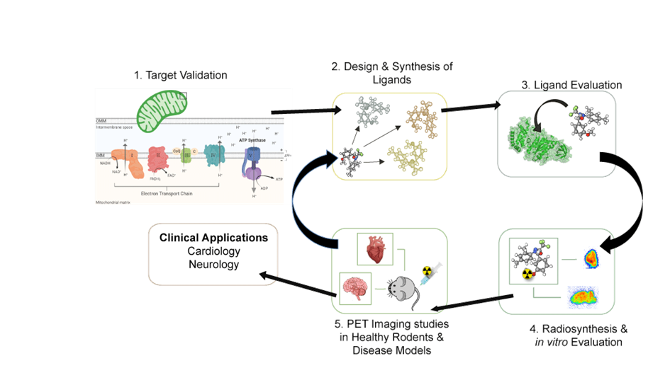
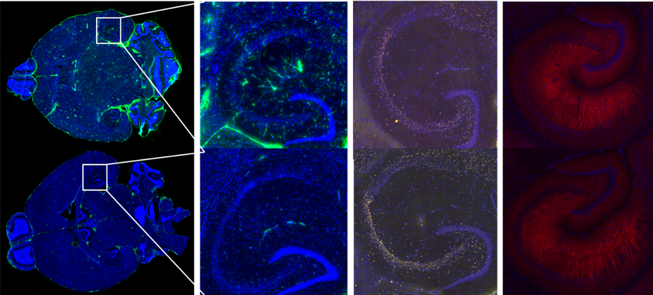
Mitochondria are fundamental organelles for metabolic homeostasis in all multicellular eukaryotes. Several mitochondrial defects have been reported in early stages of central nervous system disorders like Alzheimer’s and Parkinson’s disease and ischemic stroke (Ballinger et al., Biology and Medicine, 2005; Lin et al., Nature 2006). Therefore we are evaluating different mitochondrial biomarkers that facilitate early disease detection and monitor disease progression. The goal is to develop a suitable PET radiotracer for non-invasive imaging of mitochondrial dysfunction in humans. A series of novel radiotracer targeting mitochondrial structures have been synthesized and their in vitro characterization is currently in progress in our laboratory. In vitro evaluations include binding affinity determination, functional assays, and autoradiography. The most promising compounds will be radiolabeled and evaluated in vivo with biodistribution and PET studies using different animal models.
We perform the synthesis of new compounds, radiolabeling with Carbon-11 and Fluorine-18, in vitro binding assays, and in vivo characterization of the radiotracers in rodent models of cardiovascular and neurodegenerative disease.
This project is funded by Swiss National Science Foundation, project 205321_192409/1.